Spatial and temporal evolution of lung granulomas in a cynomolgus macaque model of Mycobacterium tuberculosis infection
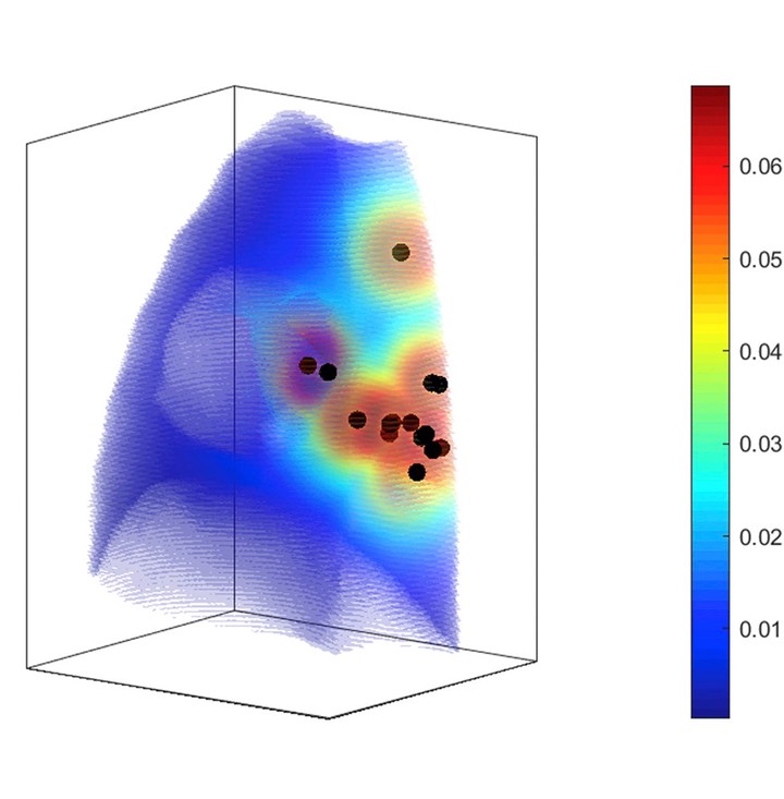 Probability of granuloma formation
Abstract
Background: Little is known about granuloma progression ofMycobacterium tuberculosisinfection in humans. Using serial positron emissiontomography and computed tomography (PET/CT) of an animal model that recapitulates human infection withM. tuberculosis,we are able totrack lung granulomas.Objective: We characterized the spatial and temporal pattern of granuloma formation during primary infection and reactivation.Methods: Serial PET/CT was performed on cynomolgus macaques (n¼28) during primary and reactivationM. tuberculosisinfection. Distancesbetween granulomas during the first six weeks post infection (“primary”granulomas) were compared to new granulomas that developed afterwards(“secondary”granulomas) using nearest neighbor analysis during primary infection, reactivation and between different routes of infection.Results: Secondary granulomas developed within 2 cm of a primary granuloma within the same lung lobe with 80% probability during the courseof primary infection, and this same pattern was observed during reactivation of latent infection after immune suppression. Using a logisticgrowth function, we were able to predict the maximum number of granulomas that would develop over the course of infection with goodcorrelation (R2¼0.96).Conclusion: These data provide important insights into the dynamic patterns of bacterial dissemination during the earliest phases of primaryinfection and reactivation tuberculosis.